Researchers in Norway aimed to determine if an inpatient rehabilitation program (IRP) was superior to an outpatient rehabilitation program (ORP) in helping women return to work following treatment for breast or gynecological cancers. Being unable to work or having to reduce work capacity due to physical and mental challenges is common after cancer treatment. Accompanying changes in quality of life and health status affect women differently and is often based upon diagnoses, treatment interventions completed, education levels and work status, according to the authors. In this article, women attending separate inpatient and outpatient locations with programs designed to reduce drop-out from work by improving physical, psychological, and social health. 51 women were included in the inpatient program, with 50 in the outpatient program. The variables assessed for outcomes included change in work status, fatigue, and health-related quality of life. At time of admission and 6 months post-admission, women ages 18-67 completed the Fatigue Questionnaire (FQ), the European Organization for the Research and Treatment of Cancer Quality of Life Questionnaire Core 30 (EORTC QLQ-C30), and information about work status.
Interventions for both groups included physical exercise, patient education, and group discussions. Educational and group discussions included topics of cancer treatment and side effects, physical activity, nutrition, work rights and return to work issues, partnership and sexuality, psychological reactions to cancer, and coping strategies. The inpatient program involved 3 weeks of stay during the week, and a 1 week follow-up 8-12 weeks later. Educational training comprised approximately 15% of time, group discussions 25% of the time, and physical activity 60% of the time. Exercise activities included Nordic walking, hiking, spinning, stretching, and relaxation. The outpatient rehabilitation occurred 5 hours/day, 1 day/week for 7 weeks. The lectures in the ORP accounted for 25% of the total time, group discussions 25% of the time, and physical activity the remaining 50% of the time. Because of the significant decrease in time spent in rehabilitation, the authors proposed that the subjects in the inpatient program would experience more significant improvements.
Fortunately, both groups improved significantly, without the expected differences in outcomes between groups. In the inpatient program, 73% of the women improved their work status compared to 76% in the outpatient program. All subjects benefited from either program in health-related quality of life and in fatigue, but no significant differences were noted between groups. One reported difference between the intervention groups is that within the inpatient group, an immediate improvement in fatigue was noted. This improvement was attributed to the 1-2 exercise sessions per day in the IRP. The authors conclude that both inpatient and outpatient rehabilitation programs for women following intervention for breast and gynecological cancers can offer substantial benefit. This conclusion is positive in that some patients may not be able to travel to participate in inpatient programs, and the cost of an outpatient program is significantly less than inpatient programs. To learn more about oncological approaches for rehabilitation, the Institute has several courses available. Susannah Haarmann's Breast Oncology course is taking place in February in Arizona. The Oncology and the Pelvic Floor A course (about the female pelvis) is instructed by Michelle Lyons and is offered next in California in May. Check the website for updates to additional oncology course dates. If you are interested in hosting a course, please contact the Institute, and if you would like to be alerted when a particular course is scheduled in your area, let the Institute know and we can keep you informed of schedule updates!
This post was written by H&W instructor Michelle Lyons, PT, MISCP, who authored and instructs the course, Special Topics in Women’s Health: Endometriosis, Infertility & Hysterectomy. She will be presenting this course this February!

Endometriosis is a common gynaecological disorder, affecting up to 15% of women of reproductive age. Because endometriosis can only be diagnosed surgically, and also because some women with the disease experience relatively minor discomfort or symptoms, there is some controversy regarding the estimates of prevalence, with some authorities stating that as many as one and three women may have endometriosis (Eskenazi & Warner 1997)
There is a wide spectrum of symptoms of endometriosis, with little or no correlation between the acuteness of the disease and the severity of the symptoms (Oliver & Overton 2014). The most commonly reported symptoms are severe dysmenorrhoea and pelvic pain between periods. Dyspareunia, dyschezia and dysuria are also commonly seen. These pain symptoms can be severe and have been reported to lead to work absences by 82% of women, with an estimated cost in Europe of €30 billion per year (EST 2005). Secondary musculoskeletal impairments caused by may include: lumbar, sacroiliac, abdominal and pelvic floor pain, muscle spasms/ myofascial trigger points, connective tissue dysfunction, urinary urgency, scar tissue adhesion and sexual dysfunction (Troyer 2007) – all of which may be responsive to skilled pelvic rehab intervention.
Endometriosis can lead to inflammation, scar tissue and adhesion formation and myofascial dysfunction throughout the abdominal and pelvic regions. This can set up a painful cycle in the pelvic floor muscles secondary to the decrease in pelvic and abdominal organ/muscle/fascia mobility which can subsequently lead to decreased circulation, tight muscles, myofascial trigger points, connective tissue dysfunction and pain and possible neural irritation.
Abdominal trigger points and pain can be commonly seen after laparascopic surgery for diagnosis or treatment. We know that fascially, the abdominal muscles are closely connected with the pelvic floor muscles and dysfunction in one group may trigger dysfunction in the other, as well as causing associated stability, postural and dynamic stability issues.
The pain created by muscle tension and dysfunction, may lead to further pain and increasing central sensitisation and further disability. Unfortunately for the endometriosis patient, as well as dealing with the problems already associated with endometriosis, she may also develop a spectrum of secondary musculo-skeletal problems, including pelvic floor dysfunction – and for some patients this may actually be responsible for the majority of their pain (Troyer 2007).
The skilled pelvic rehab therapist has much to offer this under-served patient population in terms of reducing pain and dysfunction, educating regarding self-care and exercise and helping to restore quality of life. Interested in learning more? Join me for my new course: ‘Special Topics in Women’s Health: Endometriosis, Infertility & Hysterectomy’ in San Diego this February or Chicago in June.
This post was written by H&W instructor Ginger Garner. Ginger will be presenting her Hip Labrum Injuries course in Houston in 2015!

One of the easiest ways to determine if someone is in pain is to watch the way they move. And perhaps the most commonly observed and universal movement pattern is gait. From a subtle loss of trunk rotation or pelvic translation to a gross loss of reciprocal gait, a dynamic assessment of walking is a very valuable tool in the physical therapist’s toolbox.
In evaluation of the hip, gait assessment is a critical element of the physical therapy exam. Pain-free ambulation is an essential part of measuring a person’s quality of life (QOL) and is a clinically significant functional outcome measure. Loss of hip extension and knee hyperextension prior to or at heel strike are part of several self-limiting patterns that arise from intra-articular hip injury. Dynamic gait assessment can give the therapist distinct clues as to hip pathophysiology etiology.
It was previously assumed that surgery to correct intra-articular pathology, such as in CAM-based femoracetabular impingement (FAI), would result in correction of deficiencies in gait patterning. CAM FAI limits and creates pain in the direction of hip osteokinematic flexion, adduction, and internal rotation range of motion and is caused by a lack of sphericity of the femoral head and neck, causing impingement of the labrum and/or chondral contact at the acetabulum.
A recent study published in 2013 in Gait and Posture, shows that previous assumptions about gait are incorrect. The study compared the gait of healthy controls to those with FAI and hypothesized that gait abnormalities would resolve status post surgery.
Gait measures were obtained both preoperatively and postoperatively. Researchers were surprised to find that gait abnormalities found presurgically did not automatically resolve postsurgically. Another pertinent finding is that the surgical patients not only retained their old faulty antalgic gait patterns and habits, they also adopted new abnormalities that resulted from surgical intervention, such as those arising from scar tissue, soft tissue pathology, neuromuscular patterning, or loss of arthrokinematic motion in the hip. These findings underscores the importance of early intervention via physical therapy for both operative and nonoperative patients if we want our patients to enjoy or return to a high quality of life.
Although the patients in the study who underwent FAI surgery did demonstrate decreased pain, nonoptimal preoperative gait patterns that persist postoperatively can put these patients at risk for reinjury (e.g. labral retears) or related cobmorbidities like pelvic pain, back pain, or sacroiliac joint dysfunction.
Further, a separate study published in 2009 established the presence of altered hip and pelvic biomechanics during gait, finding that those with hip FAI had decreased peak hip abduction, attenuated pelvic frontal ROM or translation, and less sagittal ROM than controls. Soft tissue restriction including scar tissue from previous or current surgeries, myofascial restriction, or neuromuscular patterning problems are, again, all important variables which must be differentially diagnosed for their possible contribution to the loss of ROM and function. Other considerations that can alter gait pattern and increase injury or reinjury risk assessment of capsular mobility, ligamentous integrity, and sacroiliac joint contributions to limited hip ROM and excursion.
To learn more about nonoperative and operative hip labral and FAI management, check out faculty member Ginger Garner's continuing education course on Extra-Articular Pelvic and Hip Labrum Injury: Differential Diagnosis and Integrative Management. The next opportunity to take the course is March of 2015 in Houston.

Research published last year in Archives of Gynecology and Obstetrics describes the benefits of "triple therapy" for symptoms of urogenital aging in postmenopausal women. The triple therapy included pelvic floor rehabilitation, intravaginal estradiol, and Lactobacillus acidophili on symptoms of urogenital atrophy, urinary tract infections (UTI's), and stress urinary incontinence (SUI) in postmenopausal women. 136 women with postmenopausal urogenital aging symptoms were divided into two groups of 68 women. Group 1 received intravaginal treatment of combined estriol (30 mcg) and Lactobacillus acidophili (50 mg) and pelvic floor rehabilitation. Group 2 received intravaginal estriol (1 mg) plus pelvic floor rehabilitation. The intravaginal treatment was applied once/day for 2 weeks and then twice/week up to 6 months.
Symptoms of urogenital aging listed by the authors include lower urinary tract issues (urinary frequency and urgency, nocturne, dysuria, recurrent UTI's, and urinary incontinence (UI)), and vaginal or vulval symptoms (vaginal dryness, itching, burning, and dyspareunia.) The connection between the microbiota Lactobacillus acidophili and vaginal health is described in the article involves the proliferation of Lactobacillus acidophili that is stimulated by estrogen. The microbiota then is reproduced in the vaginal epithelium, reduces pH, and prevents colonization of pathogens that can lead to UTI's. The pelvic floor muscle training was completed "…as explained by Castro et al…" in reference to the 2008 study assessing the efficacy of pelvic floor muscle training, vaginal cones, electrical stimulation, and no active treatment. The pelvic floor training in the Castro study included group sessions of pelvic floor muscle contractions as follows: 10 repetitions of 5 seconds contract, 5 seconds relax; 20 repetitions of 2 seconds contract, 2 seconds relax; 20 repetitions of 1 second contract, 1 second relax; 5 repetitions of 10 seconds contract, 10 seconds relax; and 5 simulated cough with strong contraction with 1 minute rest in between.
In the study, the authors assessed outcomes of urogenital symptoms in women aged 55-70 including urine cultures, colposcopic and urethral cytologic findings, urethral pressure profiles, and urethro-cystometry before and 6 months after intervention. Results included that both groups demonstrated significant improvements in symptoms and signs of urogenital atrophy , and 76% of the triple therapy group (Group 1) reported improvement in incontinence versus 41% of Group 2. Subjects in the triple therapy group also were observed to have significant improvements in colposcopic findings, urethral pressure and closure, and in abdominal pressure transmission ratio to the proximal urethra. The study concludes that combination therapy of estriol, Lactobacillus acidophili, and pelvic floor rehabilitation should be considered first-line treatment for postmenopausal symptoms of urogenital aging. To learn more about menopausal evaluation and interventions, check out faculty member Michelle Lyons' new course on Menopause: A Rehabilitation Approach. The next opportunity to take this course is in February in Orlando.
Visceral therapy is used by manual therapists, and research continues to emerge that attempts to explain the underlying mechanisms of the techniques. A study published in the Journal of Bodywork & Movement Therapies in 2012 reports on the effects of visceral therapy on pressure pain thresholds. Osteopathic visceral mobilization was applied to the sigmoid colon in 15 asymptomatic subjects. Pressure pain thresholds were measured at the L1 paraspinal muscles and 1st dorsal interossei before and after intervention. Pressure pain thresholds at the level assessed improved significantly immediately following the visceral mobilization. The effect was not found to be systemic. Hypoalgesia, therefore, may be a mechanism by which visceral mobilization affects patients who are treated with this technique.
Another research study that aimed to assess the effects of visceral manipulation (VM) on low back pain found that the addition of VM to a standard physical therapy treatment approach did not provide short term benefits. However, when the 64 patients were reassessed at 2, 6, and 52 weeks following treatment, the patients in the group with visceral manipulation were found to have less pain at 52 weeks. The patients were randomized into 2 equal groups and were provided physical therapy plus a placebo visceral treatment or a visceral treatment in addition to physical therapy. The authors propose that there may be long-term benefits of including visceral therapy in rehabilitation approaches.
If you would like to learn more about visceral techniques as well as theory and clinical application, check out the updated schedules for Ramona Horton's Visceral Mobilization 1 (VM1): The Urologic System, and Visceral Mobilization 2 (VM2): The Reproductive System. The first opportunity to take VM1 is in January in New Jersey and VM2 is scheduled in September in Ohio.
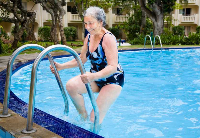
Women who are diagnosed with and treated for breast cancer commonly suffer from decreased function and fitness, and may be at risk for increased rates of functional decline than women not treated for cancer. This topic is highlighted in a paper published earlier this year in the International Journal of Physical Medicine & Rehabilitation. The authors of this article acknowledge that exercise during and following treatment for breast cancer has significant positive health effects including physical and psychosocial benefits. Cardiorespiratory and resistance training are often recommended to patients as modes of exercise that a breast cancer survivor should participate in, yet the authors raise the question about safety of the recommended exercise programs.
A literature review was completed and in this study 73 studies were included. The studies described exercise programs for patients with breast cancer during treatment and following treatment. The exercise programs within the studies varied widely, with aerobic exercise being prescribed from 10-60 minutes/session, 20-300 minutes/week, at light to vigorous intensity, and with a frequency of 1-7 days/week. The resistance-based exercises, although based on standard exercise principles, also varied dramatically in instructed parameters, according to the authors.
Other key information the authors report from the literature review is that many of the research studies were not representative of the typical patient diagnosed with breast cancer. In fact, as is described in this open-access article, women in the studies were usually younger, had less advanced disease, and in general were more well than the typical patient. For example, listed exclusion criteria in many of the studies included cardiovascular disease, diabetes, COPD, and stroke- some of the most common conditions that a woman with breast cancer also reports.
The take-home point of the article is that because many of the subjects in the exercise studies were not representative of the typical patient with breast cancer, the exercise recommendations may not be appropriate for generalization to most patients during or following treatment for breast cancer. The authors recommend future research considerations that include finding out why women choose to participate in exercise trials or choose to exercise on their own. With so many women simply being given general exercise instructions, the issue of supervised versus unsupervised exercise training, especially for women with more advanced illness should be considered.
If you would like to add more knowledge and skills to your toolbox for treating women diagnosed with breast cancer, you can join faculty member Susannah Haarmann's course Rehabilitation for the Breast Cancer Patient. In 2015 this course is currently scheduled in February in Arizona and in June in Illinois. We hope that you can join us for this specialty course!
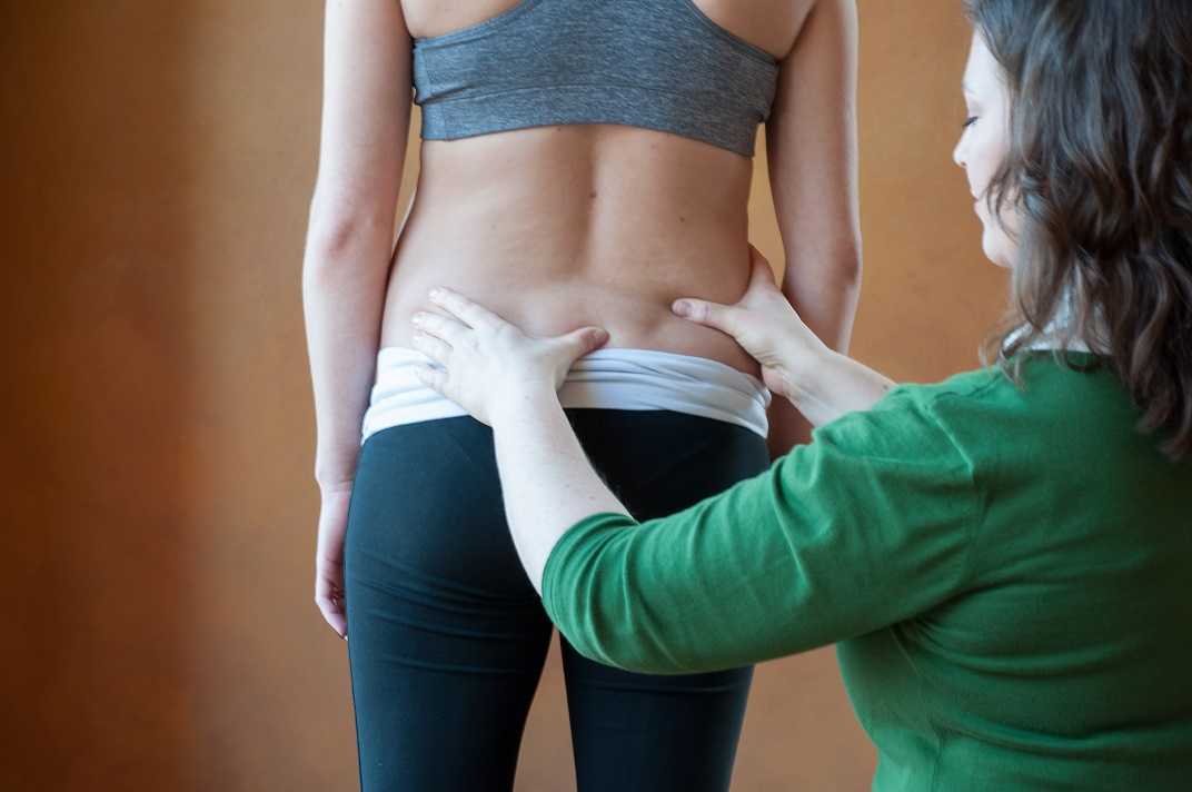
If your experience of learning sacroiliac joint mechanics, testing, and treatment has been confusing at times, trust that you are not alone in this confusion. As students have emerged from training and coursework using a variety of models to understand the joint and surrounding structures, no wonder there is disagreement and inconsistency in clinical application of learned skills. Add to this the many names for a maneuver such as the one leg standing test, and we see that the more we can streamline updated clinical knowledge and practices, the better for our profession and for our patients. I recently enjoyed reading an article summarizing assessment and treatment of sacroiliac joint (SIJ) mechanical dysfunction by Dr. Manuel Cusi, who completed a PhD thesis regarding the joint. In the article, Dr. Cusi summarized a great deal of research-based concepts related to testing and treating this issue.
Although the structure and purpose of the sacroiliac joint are described as "controversial", the author points out the foundational concept that too much or too little stability within the SIJ can create dysfunction. The "self-bracing" mechanism is provided in the pelvic girdle via both form and force closure, and Dr. Cusi points out that this joint stability that is the aim of the self-bracing mechanism must be responsive to each specific loading condition, as a function of gravity, and with coordination of muscle and ligament forces. Also according to the article, in order to assess the SIJ, the focus must be on function rather than solely on anatomic pathology.
Mechanical testing is described as being generally divided into pain provocation or palpation tests. Although we can say, based on the literature, that no one SIJ test can provide reliable data, a cluster of several tests that are positive can provide meaningful information towards a diagnosis. In order to test various aspects of SIJ function, the following tests are listed in the paradigm model. A working knowledge of the tests below, as well as pelvic joint stability tests should comprise the clinician's "toolbox" of tests for the sacroiliac joint, and this is in addition to skills used for determining other causes of SIJ pain such as disease processes or referred symptoms.
•Posterior pelvic pain provocation test (or thigh thrust)
•Long dorsal sacral ligament palpation
•Trendelenburg test
•Stork test (or Gillet test)
•Active straight leg raise (ASLR)
•Patrick's FABER and Gaenslen's test
In relation to treatment approaches, exercise training is recommended as being divided into three stages: isolation (recruiting targeted muscle in isolation of other groups), combination (muscles are recruited in various combinations to develop endurance), and function (utilizing good technique once progressed to meaningful functional tasks). While this flow of exercise training may appear very logical, the author offers that failure to progress through these three phases may be due to several factors such as poorly designed exercises that lack specificity, progressing through exercises before patient has sufficient endurance, poor adherence, and lack of appropriate exercise technique. These factors are described in the article as intrinsic to the exercise program, whereas an extrinsic factor may be failure of the exercise program to work well because of poor ligamentous stability. In this case, the author further describes the therapeutic option of prolotherapy, which will be discussed in an upcoming post.
If you are interested in learning more about the above special tests or about treatment progressions based on technique and integration, check out Peter Philip's Sacroiliac Joint and Pelvic Ring Evaluation & Treatment. The next opportunity to take the course is in January in Seattle.
Last week, Evidence in Motion, a large continuing education company that offers both live and online courses, announced that they are offering a new certificate program on pelvic rehab.
The Pelvic Health Certificate (PHC) includes a number of courses on male and female pelvic health.
At Herman & Wallace, we think it's wonderful to see yet another example of pelvic rehab being offered and promoted as part of "mainstream" physical therapy practice, particularly by an organization that has previously focused on orthopedics, manual therapy and sports medicine. The increased number of continuing education offerings on the topics of pelvic floor dysfuntion in men and women means that all the hard work our amazing faculty, course participants and certifed therapists have done over the years has finally brought pelvic rehab into the limelight!
Just like H&W, Evidence in Motion's program shys away from using "women's health" to stress that these courses cover pelvic dysfunction in men and women throughout the life cycle. While we are taking some issue with their contention "Sadly, once you are finished with school, you’ll be hard pressed to find a continuing education course that mentions the pelvic floor unless you find a course with “women’s health” in the title" (AHEM! Over here! Check out our incredible course offerings and our amazing and inspiring Certified Pelvic Rehabilitaton Practitioners!) we think it's wonderful to see yet another organization trying to get the word out to therapists, patients and referral sources about the incredible work our therapists do!
Consider the significant numbers of patients who suffer from post traumatic stress disorder, or PTSD. In the pelvic rehabilitation setting, we can think of the patients who may have experienced abuse, a loss of a child, witnessing violence, or other potential precursor. We know from the literature that there is a correlation between traumatic events such as abuse and women who have pelvic pain, within the military population, and among women who experience birth trauma. How then, can we provide patients with self-management tools to address common challenges that accompany PTSD?
Research published online this month in the journal "Mindfulness" states that in patients exposed to trauma, "…those with relatively high levels of mindfulness skills tend to have lower levels of symptoms. The article highlights previous research that has described benefits of mindfulness skills as including decreased anxiety and depression. Mindfulness-based interventions, according to the article, focuses on the development of the skills of awareness (the ability to focus on the here and now, aware of both inner and outer experiences) and acceptance (having a curious, nonjudgmental and open attitude.)
In this study, 101 patients diagnosed by a psychiatrist or psychologist with PTSD completed questionnaires including the Mini-International Neuropsychiatric Interview. The authors found skills in mindfulness correlated negatively with severity of symptoms and with "reactivity" or a tendency to respond with anxiety or depressive thoughts in response to stress. These positive relationships between mindfulness skills and symptoms of PTSD may have a significant impact on many of our patients who suffer from a variety of dysfunctions impacting health.
There is still time to sign up for Carolyn McManus's practical course on mindfulness training. Keep in mind that all therapists (not just pelvic rehab providers) will find this course immediately applicable to their caseload. Join us this November in Seattle for our new offering: Mindfulness-Based Biopsychosocial Approach to Chronic Pain.
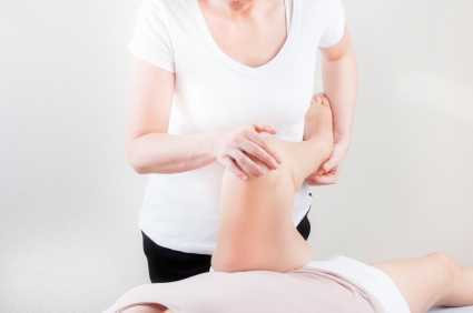
What are the roles of hip labrum innervation in both nociception and proprioception? Canadian researchers tackled this question by studying hip joints that were harvested during total hip replacement or hip resurfacing surgery. Twenty labrums were harvested and the structures in the labrum were divided into four quadrants including antero-superior (AS), postero-superior (PS), antero-inferior (AI), and postero-inferior (PI). The mean age of the subjects was near 60. The authors reported the following:
•Labral innervation is from a branch of the nerve to the quadratus femoris and the obturator nerve.
•All labrum samples had abundant free nerve endings according to the authors. These nerve endings are responsible for nociception transmission.
•Three different types of nerve end organs were noted: Vater-Pacini, Golgi-Mazonian, and Ruffini corpuscles. These nerve end organs operate to provide proprioception through their roles in pressure, deep sensation, and temperature.
•The free nerve endings and nerve end organs were observed more often in the AS and PS zones.
•The nerve endings were noted to be abundant in the superficial zones of the acetabular labrum which were for the most part avascular.
•The antero-superior zone of the labrum has abundant free nerve endings, a fact that correlates with the location of common labral tears and that also fits with the pain produced using a maneuver to test impingement in the hip (flexion, adduction, internal rotation).
•No significant differences in age was noted with respect to the innervation.
The study further states that a labral debridement may ease pain with removal of the free nerve endings, yet the labral repair may best allow for proprioceptive abilities and higher function in the injured hip. The authors also describe the findings that there are abundant myofibroblasts in the labrum that may account for the labrum's ability to heal. To learn more about healing labrums and getting patients moving, check out faculty member Ginger Garner's continuing education course on Extra-Articular Pelvic and Hip Labrum Injury: Differential Diagnosis and Integrative Management. The next opportunity to take the course is in March in Houston.
By accepting you will be accessing a service provided by a third-party external to https://hermanwallace.com./


